
DAN CARAI MAIA VIOLA 1,2,3, ALLAN SILVA ROCHA 1,2, BERNARDO LOPES CRISOSTOMO 1,2, JAIRO GRECO GARCIA 1,2, MARCELO DE TOLEDO PETRILLI 1,2, MARCOS KORUKIAN 1,2
ABSTRACT
Objective:
Evaluate risk factors related to clinical evolution and dedifferentiation of parosteal (juxtacortical) osteosarcoma to high-grade osteosarcoma.
Methods:
Retrospective cohort study performed over a period of 25 years, using data from medical records of patients diagnosed with parosteal osteosarcoma. The data were submitted to statistical analysis by Fisher’s exact test and Student’s t-test.
Results:
Of the 326 patients treated for osteosarcoma, we identified 17 patients diagnosed with parosteal osteosarcoma. Of these, 4 (23.5%) were not actually diagnosed with parosteal osteosarcoma and 4 did not have the minimum data required for analysis, being excluded from the study. Of the 9 patients studied, we observed that 3 (33.3%) evolved with tumor dedifferentiation to high-grade osteosarcoma. Moreover, 2 (66.7%) had local recurrence and 2 (66.7%) metastases.
Conclusion:
Age, sex, and the tumor size were not directly related to the dedifferentiation from parosteal osteosarcoma to high-grade osteosarcoma. The most aggressive clinical evolution – presence of local recurrences and metastasis – in parosteal osteosarcoma occurred in tumors with dedifferentiation, however, we cannot associate each other as cause and effect, but as related factors. Level of Evidence IV, Case Series.
Keywords: Bone Neoplasms, Clinical Evolution, Osteosarcoma, Juxtacortical, Recurrence, Risk Factors
INTRODUCTION
Osteosarcoma is the most common primary bone tumor, excluding hematopoietic intraosseous tumors. In its conventional form, it is a malignant tumor of high grade that produces an immature bone matrix called the osteoid. Generally, this lesion attacks the bone marrow region. 1 , 2
Tumors originated from the bone surface are 20 times less frequent and, for the most part, are of low grade. According to the World Health Organization, surface variants are parosteal steosarcoma (parosteal or juxtacortical), periosteal osteosarcoma, and high-grade surface osteosarcoma. ( 2 They correspond to 5%, 1.5%, and 0.5% of all cases of osteosarcomas. ( 2 , 3
Parosteal osteosarcoma was first described by Geschickter and Copeland in 1951 as “osteoma parosteal.” ( 2 , 4 This is a low-grade malignant tumor that is located in the metaphysis of long bones, with the distal femur (popliteal region) being the most frequent site. ( 2 , 5 Its incidence is higher in females, affecting mostly young adults between 20 and 40 years of age. ( 2 , 3 , 5 – 7
This tumor has a slow growth and may transform into a tumor with a high degree of malignancy, the dedifferentiation. ( 6 However, systemic metastases are rare. ( 6 – 8
Multiple treatment options for parosteal osteosarcoma are described, but most services opt for surgical resection of the tumor with wide margins and reconstruction with bone graft or endoprosthesis without neoadjuvant or adjuvant treatment. ( 5 – 9
Surgery performed with satisfactory margins seems to be the most important prognostic factor, since inadequate margins have been reported in association with local recurrence, dedifferentiation, and metastases, therefore, they have appeared as a negative predictor for a disease-free survival. ( 5 , 10 – 13
Dedifferentiation is reported among 8-45% of cases. It may occur as a primary event for a high-grade sarcoma (malignant fibrous histiocytoma or conventional osteosarcoma) being juxtaposed to the low-grade or secondary fibrous component after multiple recurrences of an originally low-grade tumor. ( 2 , 7 , 10 In this process there is an increase in the metastatic rate compared to conventional parosteal osteosarcoma. ( 5 , 10 – 14
Our study aims to evaluate the clinical evolution of patients diagnosed with parosteal/juxtacortical osteosarcoma and to identify probable factors related to the dedifferentiation of parosteal osteosarcoma into high-grade osteosarcoma.
METHODS
A retrospective cohort study was conducted at the Institute of Pediatric Oncology-GRAACC/UNIFESP to evaluate patients with parosteal osteosarcoma that evolved into tumor dedifferentiation to high-grade osteosarcoma. The STROBE guideline was followed for retrospective studies, Figure 1 shows the stratification of the sample. ( 15
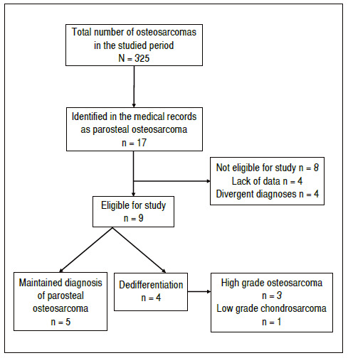
From 01/01/1993 to 31/12/2018, 17 patients diagnosed with parosteal osteosarcoma were treated in our service, which corresponded to 5.2% of the cases of osteosarcoma (326 patients). We performed a retrospective analysis of the medical records and anatomopathological reports of these patients. Of the 17 patients evaluated, 4 (23.5%) did not have a confirmed diagnosis of parosteal osteosarcoma by the anatomopathological study of the surgical specimen. Four (23.5%) patients did not present, in their medical records, the minimum data necessary for analysis. Thus, the data from nine patients were evaluated, which corresponds to our sample.
The final diagnosis of the bone tumors was considered, based on the triad of the clinical status, imaging scans, and histopathological report. ( 2 , 6 , 7 , 16 According to the institution’s routine, all cases are discussed preoperatively in a joint weekly scientific meeting between the orthopedics, oncology, radiology, and anatomopathology teams, in which the diagnosis and individual conduct of each patient are defined.
The anatomopathological analysis of all patients was performed by the same pathologist. All tests were analyzed macroscopically and microscopically, using hematoxylin-eosin staining and immunohistochemical analysis when indicated.
All patients were diagnosed with parosteal osteosarcoma after analysis of clinical data, imaging, and discussion of the biopsy result, being treated surgically for the purpose of complete tumor resection. Table 1 shows the patients’ initial diagnoses, epidemiological data, and final diagnoses of patients.
Table 1. Epidemiological data of patients with Parosteal Osteosarcoma.
| Order | Age | Sex | Initial diagnosis | Dedifferentiation | Resection size | Amputation |
|---|---|---|---|---|---|---|
| 1 | 48 | F | Parosteal osteosarcoma | – | NA* | Yes |
| 2 | 38 | F | Parosteal osteosarcoma | – | 160 mm | No |
| 3 | 34 | M | Parosteal osteosarcoma | High-grade osteosarcoma (n = 3) | 160 mm | No |
| 4 | 34 | F | Parosteal osteosarcoma | Transformation to low-grade chondrosarcoma | 340 mm | Yes |
| 5 | 25 | M | Parosteal osteosarcoma | – | 200 mm | No |
| 6 | 38 | F | Parosteal osteosarcoma | High-grade osteosarcoma (n = 3) | 200 mm | No |
| 7 | 41 | F | Parosteal osteosarcoma | – | 215 mm | Yes |
| 8 | 35 | M | Parosteal osteosarcoma | High-grade osteosarcoma (n = 3) | 200 mm | No |
| 9 | 21 | F | Parosteal osteosarcoma | – | 230 mm | No |
* Patient undergoing intralesional resection in the first procedure.
A retrospective cohort study was conducted with patients in our sample to evaluate which risk factors may be related to the evolution of dedifferentiation from osteosarcoma parosteal to high-grade osteosarcoma. Factors associated with the patients’ age at diagnosis, the presence of recurrences, and tumor size were evaluated.
We used Fisher’s exact test to describe the associations between categorical variables and the Student’s t-test to compare the means of the groups of the continuous variables. The null hypothesis (H0) adopted was that there was no difference between the means of the groups, with a significance index of 5% (p = 0.05).
Of the total of 9 patients, 3 (33.3%) were male and 6 (66.6%) were females. The mean age of the patients was 34 years (21 to 48 years). The distal femur was the segment most affected, present in 8 (88.8%) patients; and the proximal humerus was affected in one (11.1%) patient.
The research was approved by the Ethics Committee of the Institution and is registered in Plataforma Brasil under the number CAAE 28364920.9.0000.5505; opinion 3,854,662.
RESULTS
Of the 9 patients studied, we observed that 4 (44.4%) evolved due to tumor transformation. Of these, one (11.11%) patient presented transformation to low-grade chondrosarcoma and three (33.33%) presented dedifferentiation to high-grade osteosarcoma.
Of the patients in which we observed alterations in the grade of the tumor, one presented alteration of the lesion and signs of dedifferentiation while waiting for surgery. During this period, a new biopsy was submitted, which showed a change to the grade of the tumor. The patient underwent tumor resection and reconstruction with an unconventional endoprosthesis. The other two patients with dedifferentiation to high-grade osteosarcoma were submitted to systemic oncological treatment and tumor resection, according to the Brazilian Osteosarcoma Protocol. Figures 2 and 3 show imaging scans and pathological report of patient number 9 with parosteal osteosarcoma.
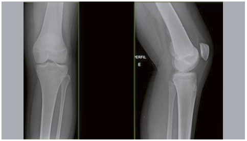
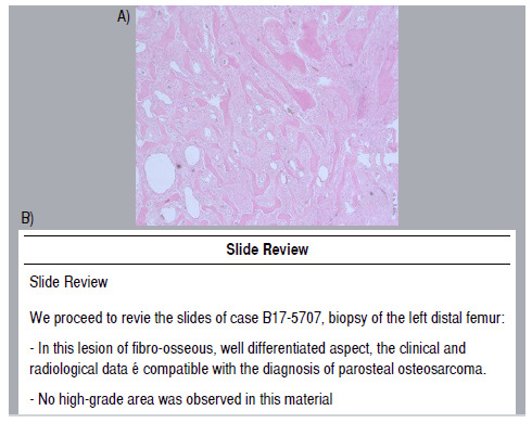
Five among the nine patients presented local recurrence of the lesion, and three patients presented dedifferentiation (two for high-grade osteosarcoma and one for low-grade chondrosarcoma) and two patients maintained the diagnosis of parosteal osteosarcoma. Four (44.5%) patients had pulmonary metastases during treatment, two patients did not present dedifferentiation, and two presented dedifferentiated. All patients underwent surgical resection of the pulmonary nodules. A fifth patient presented pulmonary nodules that were not confirmed as tumors after resection (granulomas).
One (11.1%) among the nine patients underwent intralesional surgery (curettage of lesion) after inconclusive biopsy. In the report of the anatomopathological piece, the diagnosis of parosteal osteosarcoma was evidenced, and the patient presented early recurrence in the popliteal region. A revision of the surgery was performed for resection with wide margin and reconstruction with unconventional endoprosthesis.
Three (33.3%) among the nine patients underwent limb amputation during treatment. Among them, two (22.2%) patients, after multiple approaches, evolved with periprosthesis infection and did not progress satisfactorily after a two-stage revision. The third patient underwent limb amputation after intraoperative complication due to neurovascular injury. A fourth patient presented major recurrence and ulceration in the popliteal fossa region. Amputation was indicated, but the patient did not accept treatment and, after a few months, they became deceased. Table 2 shows the surgical evolution and complications of patients.
Table 2. Surgical evolution and complications of patients.
| N° | TOPOGRAPHY | FIRST SURGERY PERFORMED | MARGINS | ORTHOPEDICS COMPLICATIONS |
|---|---|---|---|---|
| 1 | Distal femur | Intralesional curettage | Contaminated | Local recurrence, infection, implant loosening, eventual amputation |
| 2 | Distal femur | Resection + unconventional endoprosthesis | Negative | – |
| 3 | Distal femur | Resection + unconventional endoprosthesis | Negative | Local recurrence, multiple surgeries |
| 4 | Distal femur | Resection + filling with cement | Positive | Local recurrence, infection + femoral vein ligation (chronic lymphedema), amputation |
| 5 | Distal femur | Resection + unconventional endoprosthesis | Positive | – |
| 6 | Proximal humerus | Resection + unconventional endoprosthesis | Positive | – |
| 7 | Distal femur | Resection + unconventional endoprosthesis | Positive | Local recurrence, popliteal artery injury (saphenous graft) and claw toes (fibular nerve injury) |
| 8 | Distal femur | Resection + unconventional endoprosthesis | Negative | Local recurrence, early release of the implant, internal hemipelvectomy of zone II |
| 9 | Distal femur | Resection + reconstruction with plate and cement | Positive | Two revisions due to implant failure |
Regarding oncological status, in addition to the patient who deceased due to the disease, one patient with dedifferentiation is undergoing oncological treatment due to systemic recurrence. The other patients are, currently, without evidence of active disease.
Table 3 shows the analysis of the risk factors evaluated for tumor dedifferentiation (parosteal osteosarcoma and dedifferentiated high-grade osteosarcoma).
Table 3. Evaluation of risk factors for dedifferentiation into high-grade osteosarcoma.
| PAROSTEAL OSTEOSARCOMA | DEDIFFERENTIATION INTO HIGH-GRADE OSTEOSARCOMA | ||||
|---|---|---|---|---|---|
| AGE | 34.5 years (n = 6) | 35.6 years (n = 3) | p = 0.090 (t) | ||
| SEX | |||||
| F | 5 | 83.3% | 1 | 33.3% | p = 0.226 |
| M | 1 | 16.7% | 2 | 66.7% | |
| PRESENCE OF RECURRENCES | |||||
| No | 3 | 50.0% | 1 | 33.3% | P = 1.00 (F) |
| Yes | 3 | 50.0% | 2 | 66.7% | |
| MARGINS | |||||
| NEGATIVE | 1 | 16.7% | 2 | 66.7% | p = 0.226 (F) |
| POSITIVE | 5 | 83.3% | 1 | 33.3% | |
| PULMONARY METASTASES | |||||
| No | 4 | 66.6% | 1 | 33.3% | p = 0.524 (F) |
| Yes | 2 | 33.3% | 2 | 66.7% | |
| TUMOR SIZE | |||||
| Mean | 229.0 mm (n = 5) | 186.6 mm (n = 3) | p = 0.356 (t) | ||
(F): analysis by Fisher’s exact test; (t): analysis by Student’s t-test.
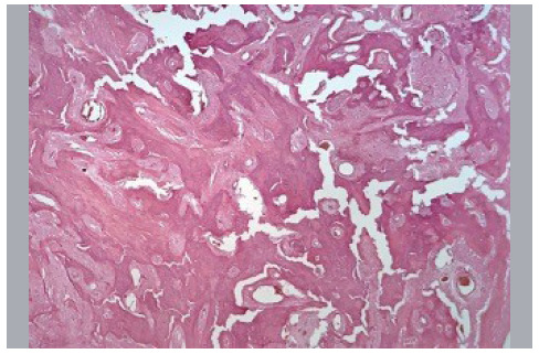
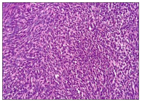
We observed that none of the factors studied showed a statistically significant association with the dedifferentiation into high-grade osteosarcoma. Figure 4 shows a photomicrographic slide of a patient with parosteal osteosarcoma, and Figure 5 shows the photomicrographic slide of the same patient after dedifferentiation to high-grade sarcoma.
DISCUSSION
Juxtacortical/parosteal osteosarcoma is an extremely rare pathology. In all publications, we found case series with few patients. Our sample is small, but we were able to carefully study each patient. We found 3 (33.33%) patients with tumor dedifferentiation, a higher number than that found in the Rizzoli Institute5,12 (24.1% and 24.6%) and in the Mayo Clinic 13 (16%), but lower than the numbers of M.D. Anderson 10 (43%). The lack of follow-up of patients may be a factor of confusion in this data. Many patients come from other regions for diagnosis or even for opinions on treatment and are registered in the medical records, but do not perform the follow-up in our service. Unlike our numbers, the Rizzoli Institute counts these data in the denominator of incidence rate. ( 5 , 12
The mean age of patients who presented dedifferentiation in the literature is slightly higher than that with parosteals osteosarcomas (35.6 years versus 34.5 years, p = 0.090). Bertoni et al. ( 12 identified a mean age of 36 years for patients with dedifferentiation. In another series of the same service, the authors found the mean age of 31 years for cases of parosteal osteosarcoma. ( 5 Sheth et al. ( 10 showed a mean lower age in dedifferentiated patients compared to non-dedifferentiated patients (31 years versus 34 years). With the current data, age does not seem to be an important diagnostic factor to differentiate these tumors. ( 5 ), ( 12
According to the observations in recent studies, the female sex and the distal region of the femur (popliteal region) are the most recurrent epidemiological characteristics in parosteal osteosarcoma. Such data are also found in cases that dedifferentiate, a fact that was confirmed in our work. ( 2 , 7 , 9 , 12 , 16
Since it is a low-grade tumor, the treatment focuses on obtaining wide margins and preserving the limb. ( 7 , 9 , 12 – 14 The most used reconstruction methods are unconventional endoprosthesis and plate and cement reconstruction. ( 2 , 4 , 5 , 7 – 11 Amputation is reserved only for cases in which negative margins cannot be achieved or due to complications of relapses. ( 11 , 13 , 16 In our sample, of the 9 patients studied, 33.3% of the patients underwent amputation. All patients showed local tumor recurrence.
A positive surgical margin is considered the main negative prognostic factor for recurrence. Several studies point to the correlation between local recurrence and dedifferentiation 5 , 9 , 12 – 14 . Our results are in line with these data, with 60% of recurrence cases related to contaminated or positive surgical margins.
We did not identify a direct correlation between positive margin and dedifferentiation. In literature, Sheth et al. ( 10 presents a large series of cases with dedifferentiation and also does not relate the alteration of the tumor degree with the oncological margin of surgery.
On the other hand, the follow-up time can mask the data of dedifferentiation. While some sample series reach up to 100 years; ( 12 , 13 in our series, the longest follow-up is of 25 years. Some patients may still differentiate during evolution. Another factor may be the bias of our service receiving only the more severe cases. Some less complex cases end up not being operated on our service and we lose the follow-up. ( 17
A third factor is that most of the margins we have are narrow, which we consider positive and not correct from an oncological point of view. However, in the case of a low-grade tumor, a narrow margin may be sufficient, in many cases, for complete resection of the lesion. This, associated with the fewer cases, may not have expressed the real significance of this factor. The margin alone is unlikely to be able to answer this question. There seems to be a biological factor, probably gene expression, that favors one or the other behavior. ( 17
Metastasis, regional or distant, is a factor suggestive of dedifferentiation, ( 5 , 10 , 14 since low-grade tumors generally have a low potential for metastatic dissemination. In patients with dedifferentiation, we found pulmonary metastases in 66% of patients, while in those without dedifferentiation this rate is 33%. Although high, these values corroborate with Sheth et al. ( 10 and Bertoni et al. ( 12 who consider that metastases are more frequent in dedifferentiated tumors.
Tumor size also does not seem to be a factor related to dedifferentiation. In our series we found that dedifferentiated tumors were smaller than non-dedifferentiated tumors (186 mm versus 229 mm). On the other hand, Lin et al. ( 18 identified larger sizes in dedifferentiated tumors, but with lower means than those found. Ruengwanichayakun et al. ( 5 and Okada et al. ( 13 found a mean size smaller than 100 mm (76 mm and 90 mm) for parosteal osteosarcomas, unrelated to survival.
Probably, the diagnosis of dedifferentiated tumors, since they are more symptomatic, occurs in a period of time prior to that of conventional parosteal osteosarcomas. This may explain the size difference we found. On the other hand, the difficulty of access to specialized health services can be represented by the difference in magnitude of tumor size when compared to those found in the literature.
The main limitation of this study is the sample size, due to the low prevalence of parosteal osteosarcoma. Thus, the statistical studies carried out are intended to support the findings and to enable a comparison between the numbers found, without intending to supply a definitive answer and exhaust the theme. Factors intrinsic to the tumor, regarding gene expression, ( 17 may better explain why some patients have dedifferentiation and others do not. This approach should also be considered for future analyses on the subject.
CONCLUSION
Parosteal osteosarcoma, when it does not dedifferentiate to a high degree, presents less aggressive clinical evolution. The ones that dedifferentiated into high-grade tumors have a natural history equivalent to conventional osteosarcoma.
We identified that the age and size of the tumor are not directly related to dedifferentiation. On the other hand, dedifferentiated tumors are related to a rate of local recurrence and higher metastasis than parosteal osteosarcomas. The theme requires studies with bigger sample and other factors related to tumor biology to more accurately identify risk factors associated with dedifferentiation and poor evolution of parosteal osteosarcoma.
ACKNOWLEDGEMENTS
We thank Prof. Dr. Maria Teresa Seixas Alves, from the Department of Pathological Anatomy of EPM/UNIFESP, for contributing to the topics of pathological anatomy and for the photomicrographs presented in this study.
Footnotes
The study was conducted at Institute of Pediatric Oncology and Department of Orthopedics and Traumatology of Paulista School of Medicine of Universidade Federal de São Paulo.
REFERENCES
- 1.Klein MJ, Siegal GP. Osteosarcoma anatomic and histologic variants. Am J Clin Pathol. 2006;125(4):555–581. doi: 10.1309/UC6K-QHLD-9LV2-KENN. [DOI] [PubMed] [Google Scholar]
- 2.Fletcher CDM, Bridge JA, Hogendoom PCW, Mertens F. WHO classification of tumours of soft tissue and bone. 4. Lyon: IARC Press; 2013. [Google Scholar]
- 3.Nouri H, Ben Maitigue M, Abid L, Nouri N, Abdelkader A, Bouaziz M, Mestiri M. Surface osteosarcoma clinical features and therapeutic implications. J Bone Oncol. 2015;4(4):115–123. doi: 10.1016/j.jbo.2015.07.002. [DOI] [PMC free article] [PubMed] [Google Scholar]
- 4.Hang JF, Chen PCH. Parosteal osteosarcoma. Arch Pathol Lab Med. 2014;138(5):694–699. doi: 10.5858/arpa.2013-0030-RS. [DOI] [PubMed] [Google Scholar]
- 5.Ruengwanichayakun P, Gambarotti M, Frisoni T, Gibertoni D, Guaraldi F, Sbaraglia M. Parosteal osteosarcoma a monocentric retrospective analysis of 195 patients. Hum Pathol. 2019;91:11–18. doi: 10.1016/j.humpath.2019.05.009. [DOI] [PubMed] [Google Scholar]
- 6.Campanacci M, Picci P, Gherlinzoni F, Guerra A, Bertoni F, Neff JR. Parosteal osteosarcoma. J Bone Joint Surg Br. 1984;66(3):313–321. doi: 10.1302/0301-620X.66B3.6586725. [DOI] [PubMed] [Google Scholar]
- 7.Jesus-Garcia R. Diagnóstico e tratamento de tumores ósseos. 2. Rio de Janeiro: Elsevier; 2013. [Google Scholar]
- 8.Nanci F, Neto, Marchiori E, Vianna AD, Aymoré IL, Almeida ALB, Irion KL, Collares FB. Parosteal osteosarcoma conventional radiology findings. Radiol Bras. 2007;40(2):81–86. [Google Scholar]
- 9.Wilke BK, Cooper AR, Gibbs CP, Scarborough MT, Spiguel AR. Long-term functional outcomes of distal femoral replacements compared to geographic resections for parosteal osteosarcomas of the distal femur. Iowa Orthop J. 2018;38:177–181. [PMC free article] [PubMed] [Google Scholar]
- 10.Sheth DS, Yasko AW, Raymond AK, Ayala AG, Carrasco CH, Benjamin RS. Conventional and dedifferentiated parosteal osteosarcoma Diagnosis, treatment, and outcome. Cancer. 1996;78(10):2136–2145. [PubMed] [Google Scholar]
- 11.Lau TW, Wong JW, Yip DK, Chien EP, Shek TW, Wong LL. Local recurrence of parosteal osteosarcoma adjacent to a prosthesis after 20 years a case report. J Orthop Surg (Hong Kong) 2004;12(2):263–266. doi: 10.1177/230949900401200225. [DOI] [PubMed] [Google Scholar]
- 12.Bertoni F, Bacchini P, Staals EL, Davidovitz P. Dedifferentiated parosteal osteosarcoma the experience of the Rizzoli Institute. Cancer. 2005;103(11):2373–2382. doi: 10.1002/cncr.21039. [DOI] [PubMed] [Google Scholar]
- 13.Okada K, Frassica FJ, Sim FH, Beabout JW, Bond JR, Unni KK. Parosteal osteosarcoma A clinicopathological study. J Bone Joint Surg Am. 1994;76(3):366–378. doi: 10.2106/00004623-199403000-00007. [DOI] [PubMed] [Google Scholar]
- 14.Laitinen M, Parry M, Albergo JI, Jeys L, Abudu A, Carter S, et al. The prognostic and therapeutic factors which influence the oncological outcome of parosteal osteosarcoma. Bone Joint J. 2015;97-B(12):1698–1703. doi: 10.1302/0301-620X.97B12.35749. [DOI] [PubMed] [Google Scholar]
- 15.von Elm E, Altman DG, Egger M, Pocock SJ, Gøtzsche PC, Vandenbroucke JP, STROBE Initiative The Strengthening the Reporting of Observational Studies in Epidemiology (STROBE) statement guidelines for reporting observational studies. Lancet. 2007;370(9596):1453–1457. doi: 10.1016/S0140-6736(07)61602-X. [DOI] [PubMed] [Google Scholar]
- 16.Enneking WF, Spanier SS, Goodman MA. Current concepts review The surgical staging of musculoskeletal sarcoma. J Bone Joint Surg Am. 1980;62(6):1027–1030. [PubMed] [Google Scholar]
- 17.He X, Pang Z, Zhang X, Lan T, Chen H, Chen M. Consistent amplification of FRS2 and MDM2 in low-grade osteosarcoma a genetic study of 22 cases with clinicopathologic analysis. Am J Surg Pathol. 2018;42(9):1143–1155. doi: 10.1097/PAS.0000000000001125. [DOI] [PubMed] [Google Scholar]
- 18.Lin HY, Wu HTH, Wu PK, Wu CL, Chen PCH, Chen WM, Guo WY. Can imaging distinguish between low-grade and dedifferentiated parosteal osteosarcoma. J Chin Med Assoc. 2018;81(10):912–919. doi: 10.1016/j.jcma.2018.01.014. [DOI] [PubMed] [Google Scholar]